Demystifying Urology Treatment: How to Navigate Diagnosis, Therapies, and Specialist Care
Introduction
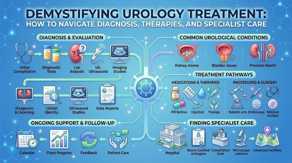
Receiving news that something is amiss with your bladder, kidneys, or prostate can feel overwhelming. From the initial moment you notice persistent discomfort or receive an unexpected lab result, you are asked to navigate unfamiliar medical language, weigh complex diagnostic options, and make significant personal healthcare choices. While modern medicine offers more therapeutic pathways than ever before, having numerous choices can sometimes add to your uncertainty. Approaching your care from an active, patient-centered perspective helps transform this uncertainty into clarity. When you understand what causes specific symptoms, how diagnostic evaluations work, and the differences between conservative monitoring and advanced surgical interventions, you can participate meaningfully in every conversation with your care team. This guide walks you through essential urological conditions, diagnostic stages, modern therapies, and the key factors to evaluate when choosing clinical centers and experienced specialists.
The Urological System from a Clinical Standpoint
The genitourinary tract functions as both an advanced waste-filtration system and an essential regulator of internal fluid balance. The kidneys continuously process the body's circulating blood volume to filter metabolic wastes, balance electrolytes, and produce urine. This fluid is transported down the muscular ureters into the bladder, which stores it until controlled release occurs via the urethra. In male anatomy, the prostate gland encircles the bladder outlet, intimately connecting urinary and reproductive function.
When disease or structural abnormalities disrupt this circuit, the effects are rarely localized. A blockage in the lower tract can rapidly generate upstream pressure that threatens delicate kidney parenchyma. Similarly, systemic metabolic shifts frequently manifest as painful crystalline formations in the renal calyces. Clinical urologists view the genitourinary tract as an interconnected mechanical and biochemical system where timely diagnostic intervention prevents permanent tissue damage and long-term functional loss.
Clinical Evaluation of Symptoms and Early Warning Indicators
In a clinical setting, patient symptoms provide initial clues that guide diagnostic testing. Because different conditions often produce identical signs, experienced physicians categorize symptoms by their underlying physiological mechanisms rather than making premature diagnostic assumptions.
Lower urinary tract voiding symptoms: Hesitancy, weak stream, intermittency, or terminal dribbling typically signal physical outflow obstruction, such as an enlarged prostate or urethral stricture.
Storage and irritative signs: Sudden urgency, daytime frequency, and multiple nocturnal awakenings often indicate bladder wall inflammation, involuntary detrusor muscle contractions, or localized bacterial irritation.
Hematuria (blood in the urine): Microscopic or gross presence of red blood cells in the urine is an objective clinical finding that always demands formal investigation to rule out stones, vascular anomalies, or urothelial malignancies.
Referred visceral pain: Severe, sharp, colicky flank pain radiating along the inguinal ligament typically points to acute ureteral obstruction, while persistent, dull suprapubic aching reflects chronic bladder distension or pelvic inflammation.
Underlying Etiology and Pathophysiological Drivers
Urological pathologies originate from a wide combination of congenital structural defects, progressive age-related degeneration, and systemic environmental influences.
Cellular and hormonal senescence: With advancing age, natural hormonal shifts drive benign cellular proliferation within the transitional zone of the prostate, narrowing the proximal urethra in the vast majority of aging men.
Urinary supersaturation and metabolic factors: Inadequate daily hydration combined with high dietary sodium, animal purines, or metabolic anomalies creates high mineral solute concentrations, prompting spontaneous crystal nucleation and stone formation.
Structural and functional stagnation: Neurogenic bladder disorders, spinal cord injuries, or anatomic strictures prevent complete bladder emptying, leaving residual pools of static urine that serve as fertile breeding grounds for persistent bacteria.
Genetic predispositions: Familial clustering strongly impacts oncological risk, particularly for early-onset prostate or clear cell renal cell carcinomas, requiring specialized screening schedules.
Diagnostic Protocols and Evidence-Based Testing
Evidence-based clinical medicine relies on a structured, phased diagnostic strategy. Physicians order specialized testing based on an initial physical examination and clinical history to minimize diagnostic delay and avoid unnecessary procedures.
Urine diagnostics: Dipstick urinalysis rapidly identifies nitrites and leukocyte esterase, microscopic examination quantifies red and white blood cells, and formal microbiological cultures identify bacterial strains alongside specific antibiotic sensitivities.
Renal and prostate serum markers: Serum creatinine and estimated glomerular filtration rate (eGFR) assess renal clearance capacity. Serum PSA is evaluated in conjunction with digital rectal examinations and age-adjusted baseline trends to assess prostate health.
Ultrasonography: Diagnostic ultrasound provides high-resolution imaging of renal parenchyma, hydro-nephrotic distension, post-void residual urine volume, and prostate geometry without ionizing radiation.
Helical computerized tomography (CT): Non-contrast thin-slice CT scans serve as the clinical gold standard for detecting and sizing urinary calculi across the entire urinary tract, while contrast-enhanced scans evaluate soft-tissue renal masses.
Endoscopic visualization (Cystoscopy): Directly passing a flexible fiber-optic or digital cystoscope through the urethra allows direct visualization of the urothelium, locating strictures, stones, and suspicious mucosal lesions.
The Continuum of Urology Treatment Approaches
Modern clinical pathways follow an escalating ladder of intervention, beginning with non-invasive measures and progressing to advanced surgical procedures only when clinically indicated.
Conservative Management and Active Surveillance
Clinical evidence shows that immediate invasive treatment is not always the best path. Watchful waiting or structured active surveillance is widely used for asymptomatic conditions and low-risk pathologies. Scheduled imaging, repeated blood panels, and clinical check-ups monitor the condition closely, avoiding procedural risks while preserving the option to intervene if the condition progresses.
Pharmacological Therapy
Targeted pharmaceuticals alter physiological processes to relieve symptoms. Clinicians use alpha-1 adrenergic antagonists to relax smooth muscle tone at the bladder neck, 5-alpha-reductase inhibitors to reduce glandular prostate volume over time, and antimuscarinic or beta-3 adrenergic agonists to stabilize an overactive detrusor muscle.
Endourological and Minimally Invasive Therapies
Endourology bypasses the need for open abdominal access by working through natural urinary channels. Using laser lithotripsy, micro-baskets, convective water vapor, or mechanical tissue retraction, specialists can resolve blockages and remove stones with minimal healthy tissue disruption and short recovery periods.
Advanced Surgical Reconstruction and Oncology
When organ preservation, extensive reconstruction, or cancer control requires direct surgical access, specialists turn to open, laparoscopic, or robotic-assisted approaches. These operations focus on oncological clearance while preserving delicate vascular and nerve structures responsible for continence and sexual function.
Clinical Breakdown of Core Urological Conditions
Kidney Stone Treatment
Renal calculi represent a mechanical disruption of urinary drainage. Clinical decisions regarding kidney stone treatment balance stone size, anatomical position, composition density (measured in Hounsfield units), and the degree of obstruction.
Medical expulsive therapy: Small stones under 5 mm located in the distal ureter often pass spontaneously with hydration, anti-inflammatory pain management, and alpha-blocker therapy.
Extracorporeal Shock Wave Lithotripsy (ESWL): For non-obstructive renal calculi under 15 mm with favorable density, acoustic sound waves generated externally fracture the stone into passable gravel.
Ureteroscopy with laser fragmentation: For ureteral or intrarenal stones resistant to external shock waves, a flexible ureteroscope coupled with a holmium or thulium laser allows direct pulverization and basket extraction under visual guidance.
Percutaneous Nephrolithotomy (PCNL): For complex stones over 20 mm, such as staghorn calculi, direct needle access through the flank provides a channel for ultrasonic or pneumatic lithotripters to fragment and clear large stone volumes.
Enlarged Prostate Treatment
Benign prostatic hyperplasia is a progressive condition requiring individualized treatment based on symptom severity, urinary flow rate, and post-void residual volumes.
Medical stabilization: Mild to moderate symptoms typically respond well to dual pharmacological therapy that relaxes smooth muscle and shrinks enlarged tissue.
Minimally invasive office procedures: Interventions such as transurethral water vapor thermal therapy or prostatic urethral lift implants offer effective symptom relief for appropriately selected prostate sizes, without requiring general anesthesia or impacting sexual function.
Surgical resective procedures: For severe obstruction or urinary retention, surgical options include monopolar or bipolar TURP, along with advanced laser procedures like Holmium Laser Enucleation of the Prostate (HoLEP), which remove the obstructing adenoma to restore an open urinary channel.
Prostate Cancer Treatment
In urologic oncology, prostate cancer care focuses on risk stratification. Physicians analyze baseline PSA levels, digital examination findings, multiparametric MRI findings, and biopsy pathology to categorize tumors as low-, intermediate-, or high-risk.
Active surveillance: For low-risk or indolent tumors, treatment involves structured surveillance protocols—including serial PSA tests, repeat MRI imaging, and periodic biopsies—to prevent overtreatment of clinically insignificant disease.
Radical prostatectomy: For localized, intermediate- or high-risk disease in patients with reasonable life expectancy, surgical removal of the prostate and seminal vesicles offers definitive local cancer control.
Radiation therapy: High-dose external beam radiation, often combined with temporary androgen deprivation therapy, or radioactive seed implants (brachytherapy) provide durable oncological control comparable to surgery in appropriate candidates.
Systemic therapies: For advanced, metastatic, or castrate-resistant disease, clinicians use androgen receptor pathway inhibitors, systemic chemotherapy, radioligand therapies, and targeted immunotherapy to control cancer spread.
Urinary Tract Infection Treatment
Urinary tract infections require clear clinical differentiation between uncomplicated lower-tract cystitis and complicated infections involving the upper tract, structural abnormalities, or systemic symptoms.
Prescribing antibiotics requires diagnostic discipline. Empirical therapy should rely on regional antibiogram patterns and be adjusted promptly once urine culture and susceptibility profiles return. For patients with recurrent infections, physicians look beyond basic bacterial exposure to evaluate anatomical abnormalities, post-void urinary retention, or bacterial biofilms on hidden calculi.
Bladder Cancer Treatment
Urothelial carcinomas require careful staging to determine whether the tumor remains superficial or has invaded the deeper muscularis propria of the bladder wall.
Transurethral Resection of Bladder Tumor (TURBT): This procedure serves both as an initial therapeutic resection of visible tumors and as the critical staging biopsy that assesses depth of invasion.
Intravesical immunotherapies: Non-muscle-invasive tumors with aggressive biological features are treated with intravesical installations, such as Bacillus Calmette-Guérin (BCG) or topical chemotherapy, to stimulate an immune response and prevent recurrence.
Radical cystectomy with urinary diversion: When cancer invades the muscular wall, the standard curative approach involves surgical removal of the bladder, nearby pelvic lymph nodes, and adjacent reproductive organs, followed by reconstructing the urinary tract using an ileal conduit or orthotopic neobladder.
Robotic Urology Surgery
Robotic-assisted surgery represents an evolutionary refinement of minimally invasive laparoscopy. Rather than operating directly with rigid, straight instruments, the surgeon controls articulating instruments through small laparoscopic ports from an ergonomic master console.
Technical capabilities: Robotic systems provide high-definition 3D visualization, up to ten-fold optical magnification, motion scaling, and tremor filtration. The instruments offer seven degrees of freedom, mimicking the articulation of the human hand in tight anatomical spaces.
Surgical applications: The platform is particularly useful for delicate reconstructive and oncologic procedures in deep pelvic or retroperitoneal spaces, such as nerve-sparing radical prostatectomies, partial nephrectomies with renal hypothermia, and complex pyeloplasties.
Clinical reality: While robotic assistance often reduces blood loss, postoperative pain, and initial hospital stays, long-term oncological and functional outcomes ultimately depend on the surgeon's anatomical knowledge and surgical volume, rather than the robotic equipment alone.
Comparative Assessment of Treatment Modalities
| Clinical Approach | Primary Indications | Key Advantages | Important Limitations |
| Active Surveillance | Low-risk prostate cancer, small non-obstructing calculi | Avoids procedural side effects; preserves baseline function | Requires strict adherence to follow-up testing protocols |
| Pharmacotherapy | Early BPH, uncomplicated UTIs, detrusor overactivity | Non-invasive; easily adjusted or discontinued | Potential drug interactions; may require ongoing use |
| Endourological Laser Surgery | Obstructive ureteral stones, non-muscle-invasive bladder tumors | Operates through natural anatomy; short recovery times | Dependent on stone size, location, and equipment availability |
| Minimally Invasive Outpatient Procedures | Mild-to-moderate BPH without severe retention | Preserves sexual function; rapid recovery without general anesthesia | Less effective for significantly enlarged prostate anatomy |
| Robotic-Assisted Laparoscopic Surgery | Localized prostate cancer, organ-confined renal cell carcinoma | Exceptional visualization; minimal blood loss; faster recovery | Highly dependent on surgeon experience; requires general anesthesia |
| Open Reconstructive Surgery | Complex anatomical trauma, multi-organ resections | Complete direct surgical access for complex repairs | Longer hospital stays and longer physical recovery timelines |
Practical Clinical Scenarios
Scenario A: Acute Obstructive Urolithiasis
A 45-year-old software architect presents to an emergency department with acute left-sided flank pain, vomiting, and microscopic hematuria. A low-dose non-contrast helical CT scan confirms an 8 mm stone impacted in the proximal ureter, causing moderate hydronephrosis.
Because the stone's size makes spontaneous passage unlikely and the obstruction impairs renal drainage, the on-call urologist advises against extended observation. After confirming the absence of systemic infection, the surgeon performs an outpatient ureteroscopy with holmium laser lithotripsy, fragments the stone into fine dust, extracts remaining fragments, and places a temporary internal double-J stent. The patient resumes normal work duties within four days, and the stent is removed uneventfully a week later.
Scenario B: Progressive Prostatic Obstruction
A 66-year-old retired teacher experiences worsening urinary hesitancy, a weak stream, and waking three times per night for over eighteen months. His American Urological Association Symptom Score is 22, indicating severe symptoms, and his post-void residual urine volume measures 160 mL.
A digital rectal examination and PSA test rule out malignancy, while transrectal ultrasound shows a 50-gram prostate without a prominent median lobe. After discussing medical side effects and surgical options, the patient chooses a minimally invasive office procedure that uses transurethral convective water vapor to ablate obstructive transition zone tissue. Six weeks post-procedure, his symptom score drops to 8, his post-void residual volume falls below 40 mL, and his nighttime awakenings decrease to once per night.
Scenario C: High-Risk Localized Prostate Oncology
A 61-year-old executive receives a routine check-up showing a PSA rise from 3.2 ng/mL to 6.8 ng/mL over two years. A multiparametric MRI reveals a 1.2 cm lesion in the peripheral zone showing restricted diffusion, with no signs of capsular extension.
A targeted MRI-ultrasound fusion biopsy confirms Gleason 4+3=7 (Grade Group 3) adenocarcinoma across three core samples. Given his relatively young age and intermediate-risk localized disease, the patient reviews options with both a radiation oncologist and a urologic surgeon. He chooses a robotic-assisted radical prostatectomy with bilateral pelvic lymphadenectomy performed at a high-volume surgical center. Surgical margins are negative, and his first postoperative PSA test confirms an undetectable level.
Evaluating Specialized Urology Hospitals and Providers
The success of complex urological care depends on matching the patient's specific condition to an experienced surgical team and a well-equipped facility. When evaluating healthcare providers, keep the following factors in mind:
Surgical subspecialization: Urology includes distinct subdisciplines, including urologic oncology, endourology, female pelvic medicine, reconstructive surgery, and male infertility. Inquire whether your provider has specific fellowship training and ongoing clinical experience in your specific condition.
Verified procedural volume: Studies consistently show a correlation between surgical volume and clinical outcomes. Centers and surgeons who perform high annual volumes of a specific procedure typically demonstrate lower complication rates, shorter operative times, and better functional preservation.
Hospital infrastructure: Advanced urological care relies on comprehensive hospital infrastructure, including modern diagnostic suites, dedicated endourological laser rooms, robotic platforms, high-resolution pathology support, and round-the-clock intensive care teams.
Multidisciplinary clinical teams: Complex oncological, reconstructive, or renal cases benefit from formal review by multidisciplinary boards that bring together urologists, medical oncologists, radiation therapists, diagnostic radiologists, and specialized pathologists.
Navigating Local Urologist Searches Effectively
When searching for a local urologist, consider logistical and operational details alongside clinical credentials:
Identify the clinical subspecialty: General urologists manage common conditions like uncomplicated stones, recurrent UTIs, and initial BPH assessments. Complex oncological resections or intricate pelvic reconstructions, however, often warrant travel to a specialized regional center.
Confirm hospital affiliations: Verify which regional hospitals the specialist works with for surgical admissions, emergency coverage, and diagnostic testing.
Assess care coordination: Review how the practice manages urgent communications, diagnostic turnaround times, post-procedure issues, and patient portal access.
Review insurance and coverage details: Check in-network coverage not only for the consulting specialist, but also for the facility, imaging centers, and anesthesia providers involved in scheduled procedures.
Practical Questions to Ask Your Urology Specialist
What specific physiological condition is causing my symptoms, and is it progressive?
What diagnostic tests are necessary right now, and which can be safely deferred?
What are the viable treatment alternatives, including active surveillance or non-invasive management?
What are the established success rates, clinical risks, and potential complications of the recommended procedure?
How does this intervention compare to newer minimally invasive or robotic techniques for my specific anatomy?
How many times have you personally performed this specific procedure over the past year?
What are the realistic recovery timelines, activity restrictions, and expected side effects?
What specific post-procedure warning signs require emergency clinical attention?
What long-term surveillance or follow-up testing schedule will be necessary after treatment?
Would you recommend seeking an independent second opinion before scheduling this surgery?
A Seven-Step Patient Decision-Making Framework
Step 1: Document symptoms objectively: Keep a detailed, multi-day record tracking voiding frequency, fluid intake, pain onset, and how symptoms affect your daily activities.
Step 2: Obtain an objective diagnostic evaluation: Complete basic laboratory tests, physical examinations, and recommended imaging to establish an accurate baseline.
Step 3: Map all therapeutic pathways: Ask your physician to outline the full range of options, from active surveillance and medications to surgery.
Step 4: Weigh clinical trade-offs: Compare the potential benefits of each option against its risks, recovery time, impact on lifestyle, and durability.
Step 5: Verify specialist and hospital credentials: Research hospital facilities, surgical technology, and your surgeon's specific procedural experience.
Step 6: Seek independent second opinions: For major surgical interventions or cancer diagnoses, consulting an outside specialist can confirm staging and ensure all reasonable pathways are considered.
Step 7: Choose your treatment path: Make an informed decision in partnership with your healthcare team, establishing clear follow-up plans and expectations.
Common Pitfalls to Avoid in Urological Care
Ignoring visible hematuria: Overlooking even a single, painless episode of blood in the urine often delays the diagnosis of treatable bladder or renal malignancies.
Accepting urinary problems as normal aging: Viewing progressive urinary hesitancy, frequency, or incontinence as an unavoidable part of aging causes patients to miss out on effective, quality-of-life-improving therapies.
Taking leftover antibiotics without a culture: Self-medicating for presumed urinary infections frequently fails to treat resistant bacteria and delays identification of the true underlying cause.
Assuming robotic surgery is always the best choice: While robotic assistance offers clear advantages for complex pelvic surgeries, traditional endourology or medical management is often safer and more effective for other conditions.
Relying entirely on hospital marketing: High-profile advertising does not always reflect clinical expertise. Look for objective provider credentials, subspecialty fellowship training, and institutional procedural volume.
Skipping long-term post-treatment follow-up: Successfully passing a stone or completing treatment for prostate disease does not eliminate the need to evaluate metabolic risks and monitor for recurrence.
Key Takeaways
Pursue timely evaluations: Diagnosing urinary issues early prevents minor anatomical or infectious problems from causing permanent kidney or bladder damage.
Hematuria requires immediate investigation: Any visible blood in the urine warrants prompt, formal clinical testing, including imaging and cystoscopic evaluation.
Match treatment to your specific anatomy: The best therapeutic choice depends on precise pathology, overall health, and personal goals—not on broad trends or one-size-fits-all recommendations.
Ask direct questions about surgical volume: Inquire about your surgeon's experience, procedural complication rates, and expected functional recovery outcomes.
Select appropriately equipped centers: Complex reconstructive procedures and oncologic resections yield better outcomes at facilities with high procedural volumes and dedicated multidisciplinary teams.
Maintain scheduled surveillance: Follow through with recommended post-procedure imaging, lab tests, and metabolic workups to protect your long-term urological health.
Frequently Asked Questions
What does a urologist treat?
A urologist is a surgical specialist trained to evaluate and manage disorders of the adrenal glands, kidneys, ureters, urinary bladder, and urethra in both men and women, along with the male reproductive system.
When should someone see a urologist?
You should see a specialist if you experience visible blood in your urine, severe flank pain, chronic pelvic discomfort, recurrent urinary infections, or progressive difficulty passing urine, or if you receive an abnormal lab result such as an elevated PSA.
How are kidney stones diagnosed?
Physicians diagnose kidney stones through clinical history, dipstick urinalysis, and microscopic evaluations, confirmed by imaging. A low-dose, non-contrast computerized tomography (CT) scan is the gold standard for defining precise stone size, location, and density.
What treatments are available for kidney stones?
Treatments range from hydration and medical expulsive therapy for small, passable stones to interventional procedures for larger ones. These include extracorporeal shock wave lithotripsy, ureteroscopy with laser fragmentation, and percutaneous nephrolithotomy.
What are common treatments for an enlarged prostate?
Treatment depends on the severity of obstruction. Options include lifestyle adjustments, daily medications to relax or shrink the prostate, minimally invasive office therapies, and endoscopic surgeries like TURP or laser enucleation to resect obstructing tissue.
How is prostate cancer treated?
Care depends on disease risk stratification, stage, and patient health. Approaches range from active surveillance for low-risk, indolent disease to radical prostatectomy, external or internal radiation therapy, and systemic hormone or targeted therapies for advanced cases.
When should urinary symptoms be evaluated?
You should seek medical attention if symptoms persist for more than a few days, interfere with daily activities, or are accompanied by fever, chills, severe back pain, or visible blood. Inability to pass urine requires immediate emergency evaluation.
What is robotic urology surgery?
Robotic surgery is a minimally invasive technique where the surgeon operates miniature articulating instruments through small incisions using a high-definition 3D console. It is especially useful for complex pelvic procedures like radical prostatectomies.
How can patients find a suitable urology hospital?
Look for facilities with accredited urology departments, advanced surgical equipment, high annual procedural volumes, low complication rates, and access to multidisciplinary oncological and reconstructive teams.
How can someone find an experienced urologist nearby?
Ask your primary care provider for referrals, check your health insurer's in-network directory, and review verified provider credentials. Look for board-certified specialists with dedicated fellowship training in the specific condition you need treated.
Conclusion
Navigating urological care starts with understanding your symptoms, securing an accurate diagnosis, and building an open dialogue with your medical team. Advances in diagnostic imaging, targeted pharmaceuticals, endourology, and robotic surgery give modern patients a wide variety of effective treatment choices. By understanding your options, researching qualified specialists, and discussing the risks and benefits of each approach, you can take control of your health and choose a care path designed for safe, durable recovery.